ECT For Depressed Adolescents With SI: New fMRI Study From China
Out on Pubmed, from investigators in China, is this study:
Alteration of Whole Brain ALFF/fALFF and Degree Centrality in Adolescents With Depression and Suicidal Ideation After Electroconvulsive Therapy: A Resting-State fMRI Study.
Front Hum Neurosci. 2021 Nov 11;15:762343. doi: 10.3389/fnhum.2021.762343. eCollection 2021.PMID: 34858155
The abstract is copied below:
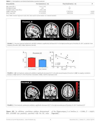
Major depressive disorder (MDD) is one of the most widespread mental disorders and can result in suicide. Suicidal ideation (SI) is strongly predictive of death by suicide, and electroconvulsive therapy (ECT) is effective for MDD, especially in patients with SI. In the present study, we aimed to determine differences in resting-state functional magnetic resonance imaging (rs-fMRI) in 14 adolescents aged 12-17 with MDD and SI at baseline and after ECT. All participants were administered the Hamilton Depression Scale (HAMD) and Beck Scale for Suicide Ideation (BSSI) and received rs-fMRI scans at baseline and after ECT. Following ECT, the amplitude of low frequency fluctuation (ALFF) and fractional ALFF (fALFF) significantly decreased in the right precentral gyrus, and the degree centrality (DC) decreased in the left triangular part of the inferior frontal gyrus and increased in the left hippocampus. There were significant negative correlations between the change of HAMD (ΔHAMD) and ALFF in the right precentral gyrus at baseline, and between the change of BSSI and the change of fALFF in the right precentral gyrus. The ΔHAMD was positively correlated with the DC value of the left hippocampus at baseline. We suggest that these brain regions may be indicators of response to ECT in adolescents with MDD and SI.
Keywords: ALFF; MDD; adolescent; degree centrality; electroconvulsive therapy; resting-state fMRI; suicidal ideation.
The pdf is here.
And from the text:
Post-ECT, our present study found changed brain function in the
precentral gyrus, hippocampus, and the triangular part of the
inferior frontal gyrus, which may indicate the mechanism of
action behind the efficacy of ECT in adolescents with MDD and SI.
This study is actually most interesting from the clinical point of view: adolescents getting treated with ECT in China, and careful monitoring of the resolution of suicidality.
Neuroimagers will want to read this paper in full, ~20 minutes, and child/adolescent psychiatrists will want to be aware of it.


Comments
Post a Comment