GEMRIC Study of Brain Structure/Function in ECT
Out on PubMed, from international investigators, is this study:

The abstract is here:
Background: Electroconvulsive therapy (ECT) is an effective treatment for severe depression and induces gray matter (GM) increases in the brain. Small-scale studies suggest that ECT also leads to changes in brain functioning, but findings are inconsistent. In this study, we investigated the influence of ECT on changes in both brain structure and function and their relation to clinical improvement using multicenter neuroimaging data from the Global ECT-MRI Research Collaboration (GEMRIC).
Methods: We analyzed T1-weighted structural magnetic resonance imaging (MRI) and functional resting-state MRI data of 88 individuals (49 male) with depressive episodes before and within one week after ECT. We performed voxel-based morphometry on the structural data and calculated fractional amplitudes of low-frequency fluctuations, regional homogeneity, degree centrality, functional connectomics, and hippocampus connectivity for the functional data in both unimodal and multimodal analyses. Longitudinal effects in the ECT group were compared to repeated measures of healthy controls (n = 27).
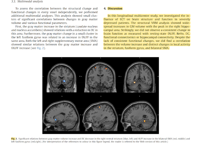
Results: Wide-spread increases in GM volume were found in patients following ECT. In contrast, no changes in any of the functional measures were observed, and there were no significant differences in structural or functional changes between ECT responders and non-responders. Multimodal analysis revealed that volume increases in the striatum, supplementary motor area and fusiform gyrus were associated with local changes in brain function.
Conclusion: These results confirm wide-spread increases in GM volume, but suggest that this is not accompanied by functional changes or associated with clinical response. Instead, focal changes in brain function appear related to individual differences in brain volume increases.
Keywords: Depression; Electroconvulsive therapy; Gray matter; Multimodal; Neuroimaging; Striatum.
And from the text:

 This study is actually more interesting for its being an international collaboration (4 GEMRIC sites) than for its findings. The lack of relationship between changes in brain structure and function/clinical outcome is disappointing, and may suggest that these particular methods of measuring brain function are inadequate.
Multimodal multi-center analysis of electroconvulsive therapy effects in depression: Brainwide gray matter increase without functional changes.
Brain Stimul. 2022 Aug 6:S1935-861X(22)00171-1. doi: 10.1016/j.brs.2022.07.053. Online ahead of print.PMID: 35944604
Background: Electroconvulsive therapy (ECT) is an effective treatment for severe depression and induces gray matter (GM) increases in the brain. Small-scale studies suggest that ECT also leads to changes in brain functioning, but findings are inconsistent. In this study, we investigated the influence of ECT on changes in both brain structure and function and their relation to clinical improvement using multicenter neuroimaging data from the Global ECT-MRI Research Collaboration (GEMRIC).
Methods: We analyzed T1-weighted structural magnetic resonance imaging (MRI) and functional resting-state MRI data of 88 individuals (49 male) with depressive episodes before and within one week after ECT. We performed voxel-based morphometry on the structural data and calculated fractional amplitudes of low-frequency fluctuations, regional homogeneity, degree centrality, functional connectomics, and hippocampus connectivity for the functional data in both unimodal and multimodal analyses. Longitudinal effects in the ECT group were compared to repeated measures of healthy controls (n = 27).
Results: Wide-spread increases in GM volume were found in patients following ECT. In contrast, no changes in any of the functional measures were observed, and there were no significant differences in structural or functional changes between ECT responders and non-responders. Multimodal analysis revealed that volume increases in the striatum, supplementary motor area and fusiform gyrus were associated with local changes in brain function.
Conclusion: These results confirm wide-spread increases in GM volume, but suggest that this is not accompanied by functional changes or associated with clinical response. Instead, focal changes in brain function appear related to individual differences in brain volume increases.
Keywords: Depression; Electroconvulsive therapy; Gray matter; Multimodal; Neuroimaging; Striatum.
The pdf is here.
Followers of the ECT neuroimaging literature will want to read this in full, ~20 minutes.


Comments
Post a Comment