MRI Textural Plasticity in Limbic Gray Matter Associated With Clinical Response to ECT for Psychosis: New Data From Korea
Out on PubMed, from researchers in Korea, is this study:
MRI textural plasticity in limbic gray matter associated with clinical response to electroconvulsive therapy for psychosis.

The abstract is copied below:
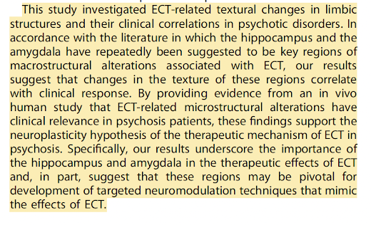
Electroconvulsive therapy (ECT) is effective against treatment-resistant psychosis, but its mechanisms remain unclear. Conventional volumetry studies have revealed plasticity in limbic structures following ECT but with inconsistent clinical relevance, as they potentially overlook subtle histological alterations. Our study analyzed microstructural changes in limbic structures after ECT using MRI texture analysis and demonstrated a correlation with clinical response. 36 schizophrenia or schizoaffective patients treated with ECT and medication, 27 patients treated with medication only, and 70 healthy controls (HCs) were included in this study. Structural MRI data were acquired before and after ECT for the ECT group and at equivalent intervals for the medication-only group. The gray matter volume and MRI texture, calculated from the gray level size zone matrix (GLSZM), were extracted from limbic structures. After normalizing texture features to HC data, group-time interactions were estimated with repeated-measures mixed models. Repeated-measures correlations between clinical variables and texture were analyzed. Volumetric group-time interactions were observed in seven of fourteen limbic structures. Group-time interactions of the normalized GLSZM large area emphasis of the left hippocampus and the right amygdala reached statistical significance. Changes in these texture features were correlated with changes in psychotic symptoms in the ECT group but not in the medication-only group. These findings provide in vivo evidence that microstructural changes in key limbic structures, hypothetically reflected by MRI texture, are associated with clinical response to ECT for psychosis. These findings support the neuroplasticity hypothesis of ECT and highlight the hippocampus and amygdala as potential targets for neuromodulation in psychosis.
The article is here.
And from the text:


Here is a very interesting neuroimaging study from Korea. It looks at "texture features" of MRI, which are purported to be reflective of the "microstructure of limbic structures." Some additional quotes from the text:
"Although not all limbic regions that exhibited volumetric
changes after ECT showed statistically significant alterations in
texture, the regions that did show significant changes in texture
features were the core brain regions that have been posited to be
the most affected by ECT...
"...one putative explanation is
that changes in the coarseness of the gray matter may reflect
reorganization of the neuroarchitecture."
"Overall, these results support the neuroplasticity hypothesis that
ECT may induce microstructural histological changes in the
human brain..."
Well, it all sounds pretty amazing.But this is a small study with significant limitations. Will it ever be replicated? And what about changes in "texture" in depression?
Kudos to these researchers, but let's see the follow up and replication.


Comments
Post a Comment